pleural effusion cat ultrasound
This form of lymphoma is often treated with chemotherapy - radiation therapy. Clinical symptoms and exam findings are usually attributed to the space occupying intrathoracic lymph nodes or secondary pleural effusion and can include dyspnea as well as noncompressible cranial thorax.
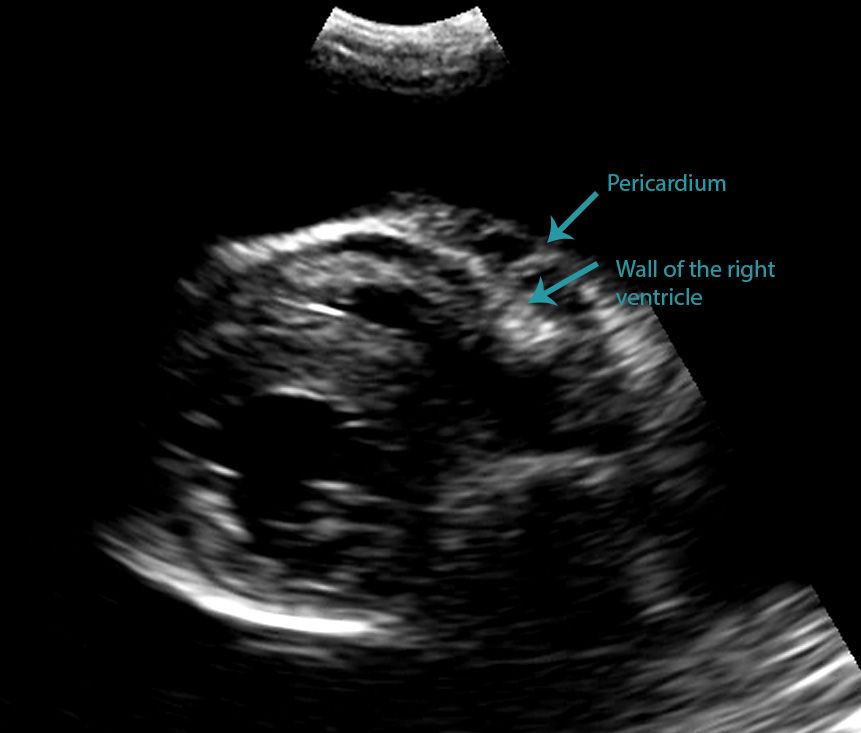
Differentiating Pericardial From Pleural Effusion Animal Ultrasound Association
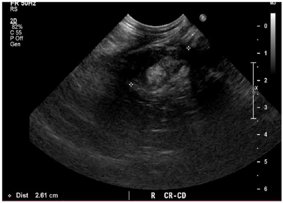
Changes noted on abdominal ultrasound examination might include renomegaly pyelectasia increased cortical or medullary echogenicity perinephric effusion hyperechoic renal medullary band hepatomegaly hypoechoic liver gallbladder abnormalities or abdominal effusion or edema.
. Pleural disease may affect the pleural space pneumothorax pleural effusion or the pleura itself thickening calcification neoplasia. Fluid in the chest space surrounding the lungs pleural effusion Bleeding into the lungs. Ethylenediamine tetra-acetic acid EDTA plasma samples are the preferred sample type for NT-proBNP testing in most clinical scenarios.
Cats with pulmonary edema or pleural effusion caused by CHF usually are presented with tachypnea and labored breathing. However pleural fluid can also be used in cats. 11 The airway detail is much improved from that of thoracic.
A company limited by guarantee. Helpful Tips to Mastering the Pedoff Probe. 4 Errors to Avoid when Measuring Aortic Valve Velocity.
Need help scheduling or have a question. He has lectured on CAT in over 40 countries across 5 continents and published over 200. You will need.
Pneumothorax Pleural Disorders pobre en hierro Anemia en español polisomnografía Estudios del sueño en español Polycythemia Vera. These changes are not specific for leptospirosis and absence of these findings does not. Computed axial tomography scan CAT scan Chest CT Scan Computed Tomography.
Trace outer edge of dense signal. For non-emergency walk-in care try an ExpressCARE location. Images such as x-rays or an ultrasound will help identify any fluid buildup foreign objects or potential tumors masses or foreign objects that may be causing the heavy breathing.
Ultrasound is useful to aid in diagnostic sampling if an isolated lesion is found on radiographs or in the presence of pleural effusion but is not useful in CCB. If heart disease is a concern once your cat is stabilized x-rays and an echocardiogram of the heart will be performed to evaluate the size and function of the heart. If your cat is having difficulty breathing this can be a life threatening emergency.
Diagnostic accuracy is higher for plasma samples than for pleural fluid samples and so plasma samples should be used in preference. How to Master Aortic Measurements with These 5. For emergency services call 911.
A service of the National Library of Medicine National Institutes of Health. Aortic Valve Anatomy. Pleurisy Pleural Effusion Pneumothorax Pleural Effusion Pleural Disorders Pleurisy Pleural Disorders Pneumonia.
Your cat remaining calm and still will have a large. Although a detailed echocardiographic examination of a cat is a difficult skill to master most clinicians will quickly learn to identify pleural effusion and. In cases of pleural effusion a thoracentesis will be performed to remove fluid from the chest which will improve breathing and provide the veterinarian with a fluid sample for analysis.
Computed tomography CT which is widely used in people with airway disease is growing in popularity for identification of canine bronchial disease as well. Depending on your cat your vet may order a mild sedative be given to your cat to potentially limit movement. Empirical diuretic treatment should be considered immediately when the index of suspicion for CHF is high for example if hypothermia and a gallop sound are present especially when echocardiography or thoracic radiography is.
Cancer Research UK is a registered charity in England and Wales 1089464 Scotland SC041666 the Isle of Man 1103 and Jersey 247. Ultrasound beam parallel to direction of flow. Review our past blogs that cover tips and techniques for measuring and obtaining the aortic valve velocity.
It is important to have your cat seen by a veterinarian as soon as possible. Nevertheless in very unstable patients with. The chest wall contains a variety of tissues including skin subcutaneous fat muscles vascular structures nerves and osseous structures vertebrae ribs costal cartilages sternum clavicles and scapulae.
Survival times tend to be longer median of nine months to a year for those. Where ultrasound is available the clinician should first establish if the dyspnoeic cat has pleural effusion andor an enlarged left atrium. High body temperature fever Diagnosis.
He has sat on CAT clinical guideline groups for National Institute for Health and Care Excellence NICE the International Society on Thrombosis and Haemostasis ISTH and Action on Smoking and Health ASH and has previously co-chaired the Scientific Sub Committee on Malignancy for ISTH. Even very stressed cats will usually tolerate ultrasound examination.
Spontaneous Cholecystopleural Fistula Leading To Biliothorax And Sepsis In A Cat

Cat Of Figure 1 Thoracic Ultrasound Revealed A Mild Hypoechoic Download Scientific Diagram

Frequency And Number Of B Lines Using A Regionally Based Lung Ultrasound Examination In Cats With Radiographically Normal Lungs Compared To Cats With Left Sided Congestive Heart Failure Lisciandro 2017 Journal

Veterinary Echocardiography Newsletter 1 Effusions Animal Ultrasound Association

Learn How To Read A Cat X Ray Long Beach Animal Hospital Radiographer Vet Medicine X Ray

Differentiating Pericardial From Pleural Effusion Animal Ultrasound Association
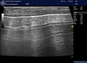
Lung Ultrasound Flooding In Fulminant Pulmonary Oedema In Cats And A Comparison With Pneumonia Vet Practice Support
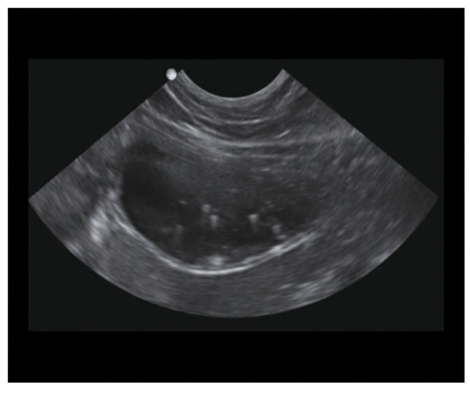
Front Line Ultrasound Imaging Of The Feline Urinary

Differentiating Pericardial From Pleural Effusion Animal Ultrasound Association